What Does a Wavefront Scan Tell Me Before Laser Eye Surgery?
Most patients coming in for a laser eye surgery assessment already know their prescription. They might tell me they are −4.00 short-sighted with some astigmatism, or that their prescription has barely changed for several years. That is useful information. But a glasses prescription describes only some of the ways an eye can bend light. There are smaller and more complex optical imperfections that ordinary glasses measurements do not describe particularly well.
A wavefront scan, or wavefront aberrometry, is one way of measuring them.
The important point is that it provides additional information.
It does not make the treatment decision by itself.
What is a wavefront measurement?
To see clearly, light entering the eye needs to be focused accurately onto the retina. In a theoretically perfect optical system, light travelling through the eye would arrive at the retina in a very orderly way. Real eyes are not perfect optical systems.
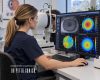
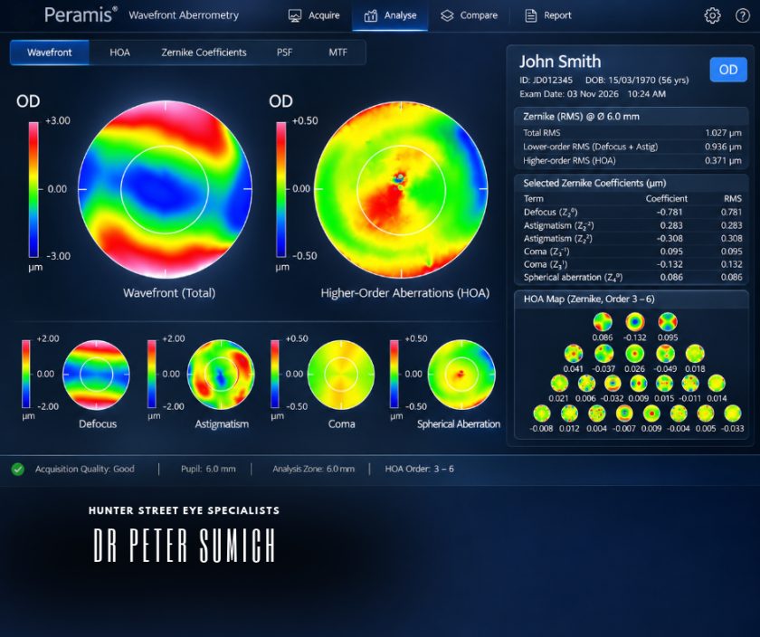
Wavefront aberrometry sends light through the eye and measures how that light is altered as it passes through the cornea, pupil, natural lens and other optical structures. The resulting measurement describes the eye’s optical aberrations. Some of those aberrations will already be familiar. Others are more complicated.
Sphere and cylinder are only the beginning
A conventional glasses prescription mainly describes what we call lower-order aberrations.
These include:
- short-sightedness
- long-sightedness
- regular astigmatism.
For most people, these account for the majority of their refractive error. But the eye can also have higher-order aberrations. These cannot usually be corrected simply by adding more sphere or cylinder to a pair of glasses.
Examples include aberrations known as coma and spherical aberration.
Patients do not need to understand the mathematics behind those terms. What matters is that higher-order aberrations can influence the quality of the image reaching the retina, even when ordinary visual acuity is good.
Why might two people with the same prescription see differently?
This is one of the useful things about wavefront testing.
Two patients can both have a prescription of −3.00 and achieve 6/6 vision on the letter chart, yet describe the quality of their vision differently. One may notice more glare around lights at night. Another may describe ghosting or smearing of images.
There are many possible reasons for symptoms like these, including the tear film, cornea, natural lens and retina.
Wavefront measurements can sometimes help us understand whether optical aberrations are contributing. That does not mean every visual symptom can be explained by an aberrometer. It gives us another piece of information.
How is this different from corneal topography?
This distinction is worth making because the tests can look similar from a patient’s point of view. Both involve sitting in front of an instrument and looking at a target. But they measure different things.
Corneal topography and tomography
Corneal imaging tells us about the shape and structure of the cornea. It can help identify irregular curvature, astigmatism, corneal thickness patterns and findings that may suggest conditions such as keratoconus. This information is particularly important when assessing whether the cornea is structurally suitable for laser treatment.
Wavefront aberrometry
Wavefront testing looks at the optical performance of the eye. The measurement includes the combined optical effect of the cornea and structures inside the eye, particularly the natural lens. The two tests therefore complement each other. One tells me more about the structure we may be treating. The other tells me more about how the optical system is behaving.
Why does it matter where an aberration comes from?
Suppose a patient has an unusual optical aberration.
The next question is: where is it coming from?
If the abnormality is clearly related to the corneal surface, corneal topography may help explain it. If the cornea looks relatively regular but the wavefront measurement remains unusual, some of the aberration may be arising from elsewhere in the eye. The natural lens can contribute to the eye’s optical aberrations, particularly as it changes with age. This becomes relevant because laser vision correction changes the cornea. It does not change an optical problem originating in the natural lens. Knowing where the problem is likely to be coming from can therefore influence how I interpret the measurements.
What is wavefront-guided laser treatment?
This is where the terminology becomes confusing.
Wavefront-guided treatment uses measurements from the individual eye’s wavefront analysis to help create a customised excimer-laser treatment profile. The aim is to address the ordinary refractive error while also considering measured higher-order aberrations. That sounds as though it must automatically be better than every alternative.
It isn’t that simple.
Most otherwise healthy eyes have relatively low levels of higher-order aberration. Evidence has not shown that wavefront-guided treatment produces universally superior standard visual outcomes for every patient compared with other modern treatment profiles.
The measurement needs to tell us something clinically useful.
Wavefront-guided and wavefront-optimised are not the same thing
These terms sound almost identical, but they mean different things.
Wavefront-guided
A wavefront-guided treatment is based on the measured optical aberrations of the individual eye.
Wavefront-optimised
A wavefront-optimised treatment does not require an individual wavefront map to customise treatment for existing higher-order aberrations. Instead, the laser profile is designed to reduce the induction of certain optical aberrations, particularly spherical aberration, during treatment. Both approaches are used in modern refractive surgery. The appropriate treatment strategy depends on the eye, the measurements and the laser platform being used.
What about topography-guided treatment?
That is another different concept.
A topography-guided treatment uses detailed information about the shape of the corneal surface to guide the treatment profile. It can be particularly relevant where corneal shape itself is contributing to irregular optics.
Wavefront-guided treatment begins with the optical aberrations measured through the eye.
Topography-guided treatment begins with the shape of the cornea.
They are related technologies, but they are not interchangeable.
The tear film matters here too
Wavefront measurements are very sensitive to the quality of the optical surface. The first surface light encounters is the tear film. If that tear film is unstable, the measured wavefront can change from one moment to the next. This is another reason I do not interpret a scan in isolation. If measurements are inconsistent, I want to understand why.
Sometimes the ocular surface needs attention before we can obtain information that is sufficiently repeatable to use for treatment planning. Current refractive-surgery guidance similarly recognises that an unstable tear film can degrade the quality of wavefront measurements.
Does everyone need wavefront-guided treatment?
No.
This is where technology can sometimes make refractive surgery sound more complicated than it needs to be. More measurements are useful when they improve our understanding of the eye. They are not useful simply because an instrument can produce them.
For one patient, wavefront information may provide clinically relevant detail about higher-order aberrations. For another patient with a straightforward prescription, regular cornea and otherwise unremarkable measurements, it may not materially change the treatment decision.
A sophisticated measurement is still only useful if we know what to do with the information.
What I am looking for
When I review wavefront information, I am not looking for a perfectly aberration-free eye.
That eye does not exist.
I am asking whether the findings are consistent with the other measurements and whether anything about the optical system changes the way I think about treatment.
- Does the wavefront result fit with the patient’s symptoms?
- Does it fit with the corneal topography?
- Are the measurements repeatable?
- Is the ocular surface stable enough for me to trust them?
And, most importantly:
Will this information change what I recommend?
If it does not, there is little value in treating the printout rather than the patient.
Measurements need to make sense together
Modern refractive assessments can produce an enormous amount of data.
- Prescription.
- Corneal curvature.
- Corneal thickness.
- Tomography.
- Pupil measurements.
- Wavefront aberrations.
- Tear-film assessment.
Each tells us something different. The skill is not collecting the largest number of measurements. It is understanding how they relate to each other.
Sometimes they tell a very consistent story. Sometimes one result does not fit and needs to be repeated or investigated. And sometimes the measurements tell us that laser treatment should not proceed.
Data is useful. Interpretation is what turns it into a clinical decision.
Frequently Asked Questions
What does a wavefront scan measure?
Wavefront aberrometry measures how light is altered as it travels through the optical system of the eye. It can quantify ordinary refractive errors as well as more complex higher-order aberrations that are not represented fully by a standard glasses prescription.
Is a wavefront scan the same as corneal topography?
No. Corneal topography primarily maps the shape of the front surface of the cornea. Wavefront aberrometry measures optical aberrations through the eye’s optical system. They provide different but complementary information.
What are higher-order aberrations?
Higher-order aberrations are more complex optical imperfections than ordinary short-sightedness, long-sightedness and regular astigmatism. Examples include coma and spherical aberration. Their clinical importance varies between patients.
Does having higher-order aberrations mean I cannot have laser eye surgery?
Not necessarily. The amount, type and likely source of the aberration need to be considered alongside corneal measurements, prescription, eye health and the proposed treatment.
Is wavefront-guided LASIK better?
Not for every patient. Wavefront-guided treatment can be useful in selected circumstances, but studies have not shown it to provide universally superior standard visual outcomes for all otherwise suitable patients compared with other modern treatment approaches.
Can dry eye affect a wavefront scan?
Yes. An unstable tear film can alter the optical surface of the eye and make wavefront measurements less consistent. Ocular-surface quality therefore needs to be considered when interpreting the result.
Is WaveScan the same as wavefront aberrometry?
“WaveScan” is sometimes used informally when discussing wavefront testing, but it is also associated with specific commercial technology. Wavefront aberrometry is the broader technical term for measuring the eye’s optical wavefront aberrations.
About the Specialist
Dr Peter Sumich is an ophthalmologist with a clinical focus on cataract surgery, laser vision correction and refractive lens exchange.
His approach to refractive treatment planning combines corneal imaging, ocular measurements, eye health and the patient’s individual visual requirements when considering whether treatment may be appropriate.
Related Articles
What I Look for in a Corneal Scan Before Laser Eye Surgery
Why corneal shape, thickness and structural stability matter before LASIK, SMILE or PRK.
Why Does the Tear Film Matter Before Laser Eye Surgery?
Why ocular-surface quality influences both diagnostic measurements and visual quality.
Does Pupil Size Matter Before Laser Eye Surgery?
How pupil measurements fit into the broader assessment of night vision and laser treatment planning.
LASIK, SMILE or PRK: Why the Right Procedure Depends on Your Eyes
Why the procedure should follow the clinical assessment rather than the other way around.
If you are considering laser vision correction, a comprehensive refractive and eye health assessment can help determine whether treatment may be appropriate and which measurements are relevant to your individual eyes.
Disclaimer
This article provides general educational information only and does not constitute individual medical advice, diagnosis or treatment. Suitability for laser vision correction, treatment selection, expected outcomes, potential benefits, limitations, alternatives and risks depend on individual circumstances and require appropriate clinical assessment.