What I Look for in a Corneal Scan Before Laser Eye Surgery
When someone comes in for a laser eye surgery assessment, they usually know their glasses prescription.
They may have been short-sighted for years, their prescription may be stable, and they may already have researched LASIK, SMILE or PRK. But a glasses prescription tells me only part of what I need to know.
Before considering laser treatment, I want to understand the structure of the cornea, the clear window at the front of the eye that we reshape during surgery. Two people can have almost identical prescriptions but very different corneas. One may be suitable for laser vision correction, while the other may not. That is why corneal imaging is such an important part of the assessment.
What the scan cannot tell us
Corneal imaging provides detailed structural information, but it does not answer every question about laser eye surgery.
Corneal imaging is only one part of the assessment. Other tests may examine the eye’s optical aberrations and how light is focused through the visual system, providing information that is different from corneal shape and thickness measurements.
It cannot tell us everything about the tear film, retinal health, natural lens or how a patient uses their vision.
What does a corneal scan show?
The cornea provides much of the eye’s focusing power. Its shape is not perfectly spherical. It has a complex curvature that varies across its surface, and those variations influence how light is focused onto the retina. Corneal imaging allows us to measure and analyse that shape in considerable detail.
Two terms patients may encounter are corneal topography and corneal tomography.
Corneal topography
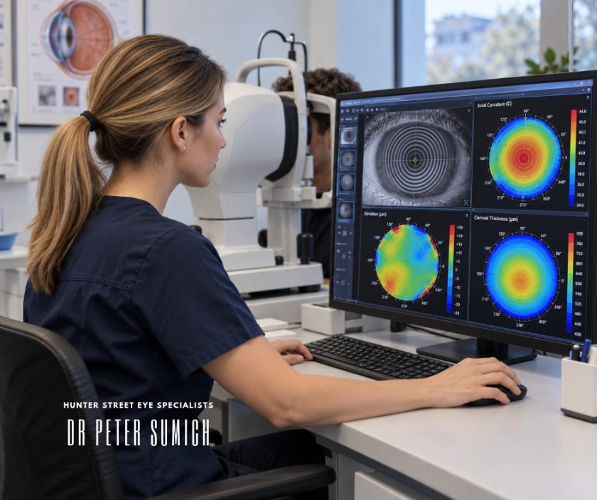
Corneal topography maps the curvature of the front surface of the cornea. The results are commonly displayed as colour-coded maps showing areas of different curvature. These maps can help identify regular astigmatism, differences between the two eyes and patterns of corneal irregularity.
Corneal tomography
Corneal tomography provides additional information about the cornea’s three-dimensional structure. Depending on the imaging system, this includes measurements of the front and back surfaces of the cornea, together with a map showing how its thickness varies. This is important because some early structural abnormalities may not be obvious from the front surface alone.
Why corneal shape matters
When I review a corneal scan, I am not simply looking for a particular number. I am looking at the overall pattern. Is the curvature reasonably regular? Are the two eyes similar? Is there an area of unusual steepening? Does the thickness distribution make sense alongside the shape?
A patient can have excellent corrected vision with glasses and still have subtle corneal abnormalities. One condition we specifically consider is keratoconus. Keratoconus is a condition in which the cornea progressively changes shape, often becoming thinner and more irregular. In its early stages, the patient may have few symptoms, and the condition may not be obvious during a routine glasses examination. This matters because laser vision correction changes the structure of the cornea. Performing refractive surgery on a cornea with an underlying tendency to become unstable can increase the risk of further structural problems.
An abnormal scan does not automatically mean someone has keratoconus. It does mean the findings need to be understood before treatment is considered.
Thickness is important, but it is not the whole story
Patients sometimes ask whether their corneas are thick enough for laser eye surgery. It is a reasonable question, but thickness alone does not establish suitability.
Laser vision correction changes the cornea’s refractive power by altering its tissue. The amount of tissue affected depends on the prescription and the procedure being considered. A higher correction may require a greater change to the cornea than a smaller correction. I therefore need to consider the original corneal thickness, the proposed treatment and how much structurally important tissue would remain. But a relatively thick cornea can still have an abnormal shape or other concerning characteristics. Equally, a thinner cornea is not automatically abnormal.
The relationship between shape, thickness and the proposed treatment is more informative than any single measurement.
Why structural stability matters
The cornea needs to maintain its shape after surgery. One uncommon but important complication of corneal refractive surgery is corneal ectasia. This occurs when the cornea progressively weakens and changes shape, potentially causing increasing short-sightedness, irregular astigmatism and reduced visual quality. Ectasia can occur after different forms of corneal refractive surgery, although the risks and structural effects are not identical between procedures. Pre-operative screening is intended to identify patients who may be at increased risk.
Corneal imaging is a major part of that process, but it is not the only consideration. Age, prescription, corneal thickness, the amount of tissue affected by treatment and other clinical findings also contribute to the assessment. No screening process can eliminate every possible risk.
Why I sometimes want measurements repeated
A scan is only useful if the measurements are reliable. The tear film can influence the quality of corneal imaging. Contact lens wear can also temporarily alter corneal shape. For that reason, patients may be asked to stop wearing contact lenses for an appropriate period before assessment. The required interval varies according to the type of lens and the individual eye. Sometimes a scan needs to be repeated because the measurements are inconsistent or the corneal surface needs further attention. If the findings remain unusual, additional testing or observation over time may be appropriate.
I would rather understand why measurements are inconsistent than make a treatment decision from a result that may not accurately represent the cornea.
Does an abnormal scan mean laser eye surgery is impossible?
Not necessarily, but it may change the recommendation.
Some irregular findings are related to measurement quality or temporary changes in the ocular surface. Others may indicate an underlying corneal condition that makes elective laser treatment inappropriate. The important distinction is whether the abnormality can be explained and whether the cornea can reasonably be expected to tolerate the proposed procedure.
Changing from LASIK to PRK or SMILE does not automatically resolve every concern about corneal stability. If the underlying cornea is unsuitable, choosing a different laser technique may not remove the risk.
In some circumstances, glasses or contact lenses remain the more appropriate option. Other refractive approaches may be considered depending on the patient’s age, prescription and eye health.
What the scan cannot tell us
Corneal imaging provides detailed structural information, but it does not answer every question about laser eye surgery. It cannot tell us everything about the tear film, retinal health, natural lens or how a patient uses their vision. Nor can a normal-looking scan guarantee that complications will not occur.
The results need to be interpreted alongside the prescription, clinical examination, medical history and the patient’s visual requirements. This is particularly important when deciding between LASIK, SMILE and PRK.
The procedure should follow the assessment, rather than the assessment being used to justify a procedure already chosen.
The value of understanding the eye before changing it
Laser vision correction is intended to change the way the eye focuses light. Before making that change, we need to understand the structure we are treating.
A corneal scan can reveal information that would not be apparent from the glasses prescription alone. Sometimes it confirms that laser treatment may be reasonable to consider. Sometimes it raises questions requiring further investigation. Occasionally, it provides a reason not to proceed.
The purpose of the assessment is not simply to determine whether a laser can correct the prescription. It is to determine whether changing the cornea is appropriate for that particular eye.
That is a distinction worth understanding before deciding on surgery.
Frequently Asked Questions
What is corneal topography?
Corneal topography is a diagnostic test that maps the curvature of the front surface of the cornea. It helps identify astigmatism, irregular curvature patterns and other features relevant to laser vision correction.
What is the difference between corneal topography and tomography?
Topography primarily measures the curvature of the front corneal surface. Tomography provides additional three-dimensional information, including the back surface and distribution of corneal thickness. Both can contribute to refractive surgery assessment.
Can I have LASIK if I have thin corneas?
Possibly, but suitability cannot be determined by thickness alone. The prescription, corneal shape, proposed treatment and amount of tissue that would remain all need to be considered. Some patients with thin corneas may not be suitable for corneal laser surgery.
Can a corneal scan detect keratoconus?
Corneal topography and tomography are important tools for detecting keratoconus, including subtle changes that may not be apparent during a routine examination. Diagnosis requires interpretation of the imaging alongside other clinical findings.
Does a normal corneal scan guarantee laser eye surgery is safe?
No. Normal imaging is reassuring but does not eliminate surgical risks. A comprehensive assessment considers multiple factors, and complications can occur even after appropriate screening.
Why do I need to stop wearing contact lenses before my assessment?
Contact lenses can temporarily influence corneal shape and affect the accuracy of measurements. The time required without lenses depends on the lens type and individual circumstances. Your treating clinic will provide instructions.
If my corneal scan is abnormal, can I have PRK instead of LASIK?
Not necessarily. Although PRK and LASIK affect the cornea differently, both change corneal tissue. An underlying condition that increases the risk of corneal instability may make several forms of laser vision correction inappropriate.
About the Specialist
Dr Peter Sumich is an ophthalmologist with a clinical focus on cataract surgery, laser vision correction and refractive lens exchange.
His approach to refractive treatment planning considers detailed ocular measurements, corneal health, prescription stability and the patient’s individual visual requirements when assessing treatment options.
Related Articles
Does Pupil Size Matter Before Laser Eye Surgery?
How pupil measurements relate to night vision, optical zones and laser treatment planning.
Why Does the Tear Film Matter Before Laser Eye Surgery?
Why the ocular surface influences the accuracy of measurements and the quality of vision.
LASIK, SMILE or PRK: Why the Right Procedure Depends on Your Eyes
How corneal anatomy, prescription and other clinical findings influence the choice of laser procedure.
If you are considering laser vision correction, a comprehensive refractive assessment can help determine whether your cornea is suitable for treatment and which options may be appropriate for your eyes.
Disclaimer
This article provides general educational information only and does not constitute individual medical advice, diagnosis or treatment. Suitability for laser vision correction, potential benefits, limitations, alternatives and risks depend on individual circumstances and require appropriate clinical assessment.